Gynecological & Pelvic Ultrasound
- High-definition Transvaginal (TVS) & pelvic ultrasound diagnostics
- 3D/4D uterine cavity mapping for fibroids, polyps & adenomyosis
- Accurate differentiation of ovarian cysts and endometriomas
- Direct scan interpretation and clinical guidance by Dr. Noopur Bajpai
Introduction
Gynecological ultrasound is the primary, non-invasive imaging modality used to evaluate the female pelvic organs, including the uterus, cervix, fallopian tubes, and ovaries. High-resolution ultrasound imaging enables rapid, accurate diagnosis of underlying pelvic pain, abnormal uterine bleeding, menstrual irregularities, and fertility hurdles without any exposure to ionizing radiation.
At Florentine IVF and Women Wellness Centre in Lucknow, ultrasound examinations are not outsourced to external diagnostic centers where clinical context is missing. Instead, Dr. Noopur Bajpai (MS Obs & Gynae, F.MAS) directly reviews or conducts high-resolution 3D/4D pelvic sonography during your consultation, correlating scan findings instantly with your clinical symptoms to design an immediate, targeted treatment plan.
Specialized Pelvic Ultrasound Modalities
We provide tailored ultrasound techniques depending on your diagnostic needs:
1. Transvaginal Sonography (TVS): The gold standard for reproductive imaging. By placing a slender ultrasound probe close to the pelvic organs, TVS provides crystal-clear resolution of the uterine wall, junctional zone, endometrial lining, and microscopic ovarian structures that abdominal ultrasound cannot match.
2. 3D/4D Uterine Cavity Reconstruction: Creates multiplanar coronal views of the uterus, allowing clear diagnosis of Mullerian duct anomalies (such as bicornuate, arcuate, or septate uterus) and distinguishing between intramural fibroids and deep adenomyosis.
3. Pelvic Color Doppler: Evaluates uterine artery blood flow and resistance indices, assessing endometrial vascular receptivity for embryo implantation and monitoring abnormal blood flow around pelvic masses.
4. Sonosalpingography (SSG / saline infusion sonography): A gentle, painless procedure infusing sterile saline to visualize the internal uterine cavity and confirm fallopian tube patency under direct real-time ultrasound imaging.
Conditions Accurately Detected
Our ultrasound diagnostic protocols identify a wide spectrum of gynecological conditions:
-
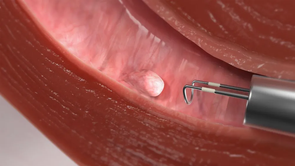
Uterine Fibroids & Polyps: Precise size, number, and location (submucosal, intramural, subserosal) to plan fertility-preserving removal.
-
Ovarian Cysts & Endometriomas: Distinguishing benign functional cysts from "chocolate" endometriotic cysts, dermoid cysts, or suspicious ovarian neoplasms.
-
Adenomyosis: Identifying uterine enlargement, asymmetric myometrial thickening, and myometrial cysts causing severe dysmenorrhea.
-
PCOS Morphology: Documenting the classic "pearl-string" arrangement of multiple peripheral follicles and increased stromal density.